
Foot drop is a frequently seen sign of various neurological and orthopedic conditions affecting the lower leg. Typically, the peroneal nerve is involved, resulting in an inability to elevate the foot or toes off the ground. This may result in a person dragging their foot when walking, increasing their danger of tripping or falling, or altering their steppage stride.
The following tests are essential and are used by doctors to assess whether you have foot drop, also known as drop foot, and if additional study or diagnosis by physicians is necessary.


Foot drop is a term that refers to having difficulties elevating the front half of your foot. It is characterized as an unusual dorsiflexion weakness of the ankle.
Heel Walk Examination
During this test, individuals have to walk on their heels for roughly 10 to 15 feet. To ensure safety, have the individual grasp a countertop or use a device such as a cane to maintain balance and stability. It is expected from the subject to struggle to start the test, or be unable to keep their afflicted foot in the air for the test duration.
If your subject’s equilibrium is off, do not try this test. A version is to stand on their heels at a counter and attempt to keep their toes off the ground. Keep an eye on the injured foot to ensure it does not come off the ground or return gently to the ground.
Braces or splints may be necessary. An afo brace around your ankle and foot, or a splint that fits inside your shoe, may assist in maintaining a normal posture for your foot.
Imaging examination
Foot drop may occur due to excessive bone development in the spinal canal or as a result of a tumor or cyst pushing on a nerve in the knee or spine. Imaging examinations may assist in identifying these sorts of issues.
- Simple X-rays use a modest dose of radiation to see a soft tissue tumor or a bone lesion that may be causing your symptoms.
- Ultrasound creates pictures of internal structures using sound waves, may check for cysts or tumors on the nerve or demonstrate edema caused by compression.
- Magnetic resonance imaging (MRI) creates detailed pictures using radio waves and a strong magnetic field. MRI is perfect for visualizing abnormalities in the soft tissue that may compress a nerve.
Jump Examination
Jump examination requires a high level of safety awareness and balance. If the patient has significant fall risk, this test should be avoided. Kindly do not attempt to finish this exam on your own. Conduct the test with the assistance of a therapist or clinician. First, ask the subject to leap off the ground while hanging onto a counter for support. Ensure that you promote getting off the ground. If the person cannot do so, this is positive evidence of foot drop.
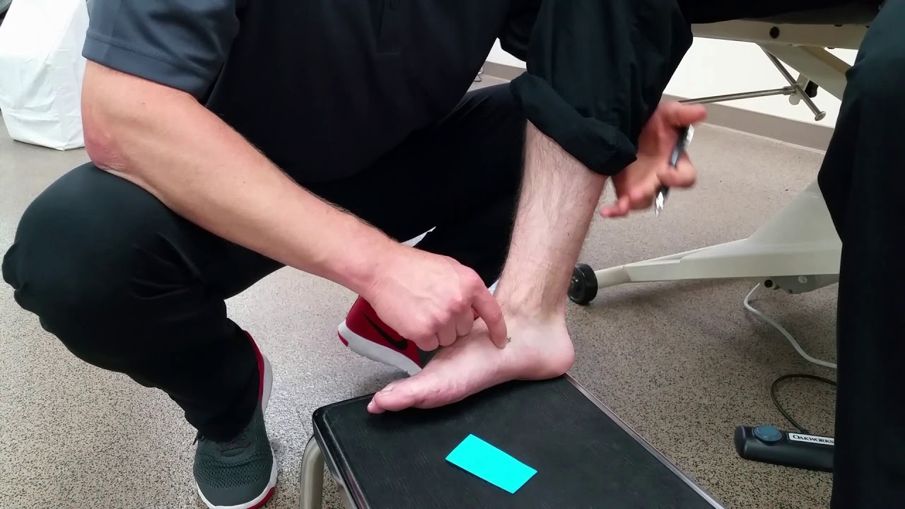
Muscle Manual Test
The manual muscle test is the third exam. This examination is referred to as a manual muscle (strength) test or MMT. This test may be conducted with or without the assistance of a doctor. However, if you are unable to reach your toes, have impaired sitting balance, or are alone, you may find that utilizing the handle of a cane assists you in completing this test.
To do this test, sit on a chair with your afflicted leg out in front of you. Begin by attempting to raise the toes and soles of your feet off the ground. If you can extend the foot off the ground, aid the foot in completing its range of motion by bringing the toes up toward the shin. Attempt to keep it there.
If the maneuver is not adequately met, this is a favorable indicator for foot drop. If you can maintain that posture for the foot, apply pressure to the top of the foot, assessing if the foot requires moderate or less force to break the holding position. In other words, this is an indication of vulnerability.
Nerve examinations
Electromyography (EMG) and nerve conduction tests measure electrical activity in muscles and nerves. Tests may be unpleasant, but they are necessary for detecting the location of the damage along the afflicted nerve.
So, these were some easy tests that can be used to diagnose foot drop.


